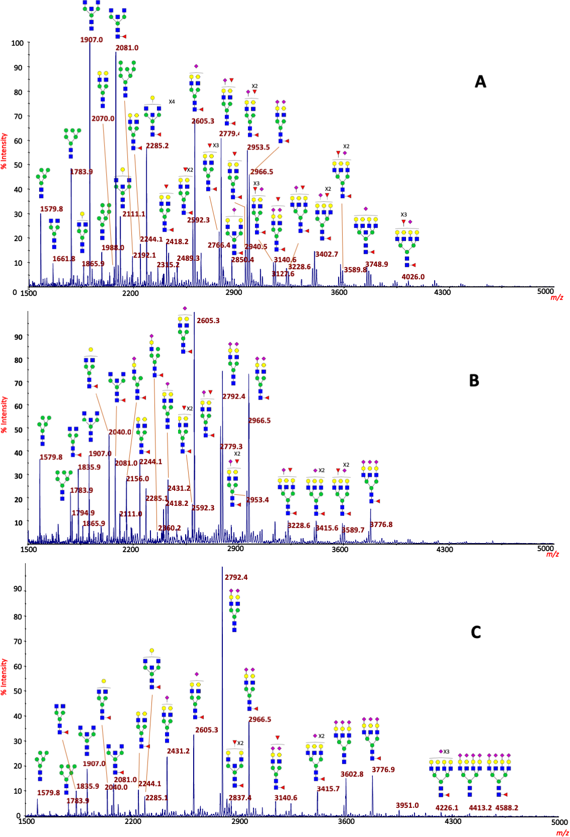
Purpose Tear fluid N-Glycome from patients affected with vernal (VKC) and atopic keratoconjunctivitis (AKC) was investigated to identify specific changes in tears and to recognize possible glyco-biomarkers. Methods The analysis of N-glycans was performed using matrix-assisted laser desorption ionization mass spectrometry on single tear samples. Tears from control normal subjects (CTRL), VKC and AKC patients were processed and treated with peptide N-glycosidase F (PNGase F) to deglycosylate N-glycoproteins. Released N-glycans were purified, permethylated and analyzed by Matrix-Assisted Laser Desorption/Ionization-Time Of Flight Mass Spectrometry and tandem Mass Spectrometry (MALDI-TOF MS and MALDI-TOF MS/MS). Results More than 150 complex N-glycans, including highly fucosylated biantennary, triantennary, tetraantennary and bisecting species, were observed in our spectra. Three distinct patterns for CTRL, VKC and AKC patients were identified in terms of relative intensities for some N-glycans structures. Major variations involved bisecting and hyperfucosylated glycoforms. The most intense ions were associated to species at m/z 1907.0 (asialo, agalacto, bisected, biantennary structure – NGA2B) in CTRL MS profiles, at m/z 2605.3 and 2966.5 in VKC, and at m/z 2792.4 in AKC corresponding to a well-known biantennary, disialylated N-glycan. Several peaks were associated to structures bearing one or two Lewis X epitopes. Structures were confirmed by MS/MS analysis. Quantitative differences among the three groups were statistically significant. Conclusions Tear MS profiles are rich in specific glycoforms, particularly those with a high fucosylation degree, indicating both core and peripheral decoration. Tear N-glycome analysis provided important information for a better comprehension of VKC and AKC alterations at the molecular level