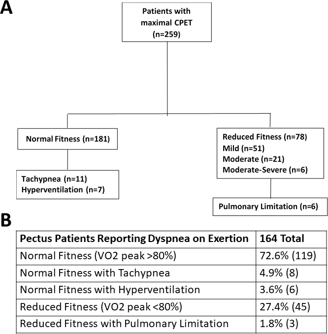
Pulmonary defects are reported in pectus excavatum but the physiological impact on exercise capacity is unclear. To test the hypothesis that pectus deformities are associated with a pulmonary impairment during exercise we performed a retrospective review on pectus patients in our center who completed a symptom questionnaire, cardiopulmonary exercise test, pulmonary function tests (PFT), and chest magnetic resonance imaging. Of 259 patients studied, dyspnea on exertion and chest pain was reported in 64% and 41% respectively. Peak oxygen uptake (VO2) was reduced in 30% and classified as mild in two-thirds. A pulmonary limitation during exercise was identified in less than 3%. Ventilatory limitations on PFT was found in 26% and classified as mild in 85%. Obstruction was the most common abnormal pattern (11%) followed by a nonspecific ventilatory limitation and restrictive pattern (7% each). There were no differences between patients with normal or abnormal PFT patterns for the anatomic degree of pectus malformation, VO2, or percentage reporting dyspnea or chest pain. Scatter plots demonstrated significant inverse relationships between severity of the pectus deformity with lung volumes on PFT and VO2 but no correlation between the severity of the pectus deformity and lung volumes during exercise. We conclude that resting lung volume measurements were associated with the anatomic degree of pectus severity but respiratory limitations during maximal exercise are uncommon and PFT patterns have poor correlation with symptomatology or VO2. These findings suggest non-respiratory causes are more likely for the high rates of dyspnea and reduced aerobic fitness reported in pectus.