Abstract Rationale: Recent studies suggest that both hypo- and hyper-inflammatory ARDS phenotypes characterize severe COVID-19-related pneumonia. The role of lung SARS-CoV-2 viral load in contributing to these phenotypes remains unknown. Objectives: To redefine COVID-19 ARDS phenotypes when considering semi-quantitative SARS-CoV-2 RT-PCR in the bronchoalveolar lavage of intubated patients. To compare the relevance of deep respiratory samples vs plasma in linking the immune response and the semi-quantitative viral loads. Methods: Eligible subjects were adults diagnosed with COVID-19 ARDS who required mechanical ventilation and underwent bronchoscopy. We recorded the immune response in the bronchoalveolar lavage and plasma and semi-quantitative SARS-CoV-2 RT-PCR in the bronchoalveolar lavage. Hierarchical clustering on principal components was applied separately on the two compartments datasets. Baseline characteristics were compared between clusters. Measurements and Results: 20 subjects were enrolled between August 2020 and March 2021. Subjects underwent bronchoscopy on average 3.6 days after intubation. All subjects were treated with dexamethasone prior to bronchoscopy, 11 of 20 (55.6%) received remdesivir and 1 of 20 (5%) received tocilizumab. Adding viral load information to the classic two cluster model of ARDS revealed a new cluster characterized by hypo-inflammatory responses and high viral load in 23.1% of the cohort. Hyperinflammatory ARDS was noted in 15.4% of subjects. Bronchoalveolar lavage clusters were more stable compared to plasma. Conclusions: We identified a unique group of critically ill subjects with COVID-19 ARDS who exhibit hypo-inflammatory responses but high viral loads in the lower airways. Our approach adds the infection dimension to ARDS phenotypes described in COVID-19 pneumonia

Matthew Plassmeyer

and 20 more

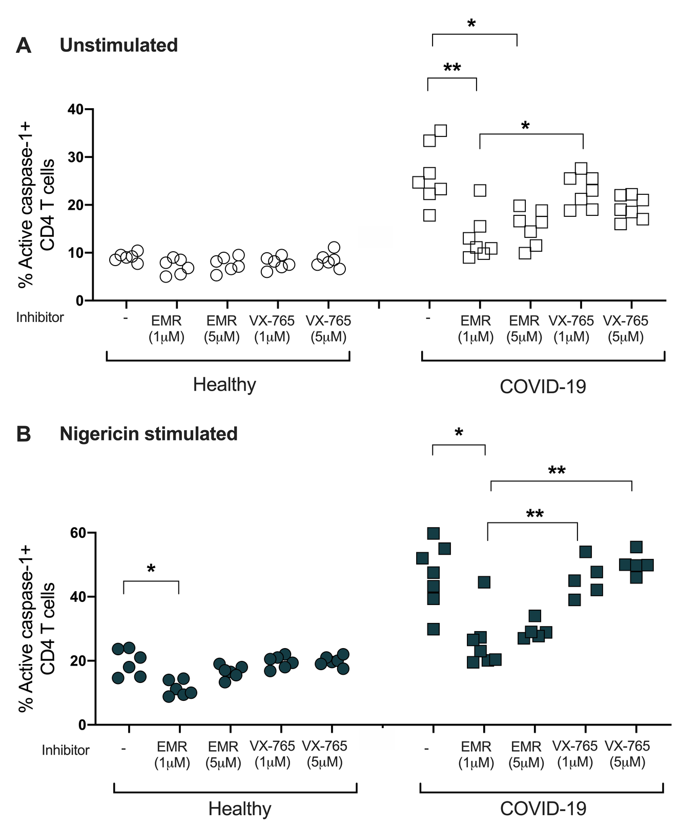
COVID-19 can present with lymphopenia and extraordinary complex multi-organ pathologies that can trigger long-term sequela. Given that inflammasome products, like caspase-1, play a role in the pathophysiology of a number of co-morbid conditions, we investigated caspases across the spectrum of COVID-19 disease. We assessed transcriptional states of multiple caspases and using flow cytometry, the expression of active caspase-1 in blood cells from COVID-19 patients in acute and convalescent stages of disease. Non-COVID-19 subjects presenting with various co-morbid conditions served as controls. Single-cell RNA-seq data of immune cells from COVID-19 patients showed a distinct caspase expression pattern in T cells, neutrophils, dendritic cells and eosinophils compared to controls. Caspase-1 was upregulated in CD4+ T-cells from hospitalized COVID-19 patients compared to unexposed controls. Post-COVID-19 patients with lingering symptoms (long-haulers) also showed up-regulated caspase-1 activity in CD4+ T-cells that ex vivo was attenuated with a select pan-caspase inhibitor. We observed elevated caspase-3/7 levels in red blood cells from COVID-19 patients compared to controls that was reduced following caspase inhibition. Our preliminary results suggest an exuberant caspase response in COVID-19 that may facilitate immune-related pathological processes leading to severe outcomes. Further clinical correlations of caspase expression in different stages of COVID-19 will be needed. Pan-caspase inhibition could emerge as a therapeutic strategy to ameliorate or prevent severe COVID-19.