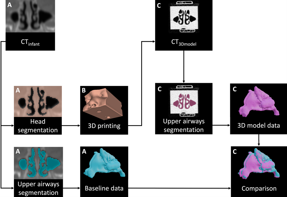
BACKGROUND: Three-dimensional (3D) printing has become increasingly affordable. Several research projects used 3D printing to create in vitro upper airways model. However, studies using a mainstream desktop 3D printer never performed geometric validation of their model. The aim of this study was to perform geometric validation of a pediatric upper airways model printed with a mainstream desktop 3D printer. METHODS: Head computerized tomography (CT) scan of a 10-month-old female underwent segmentation between airways and surrounding anatomical structures. Airways segmentation allowed their measurement for further comparison with printed model. Head segmentation enabled the creation of a 3D printable volume file. To proceed to the geometric validation of the head model, the latter underwent a CT scan. Similar segmentation work was performed on the printed model for comparison. Overlap proportion between the original infant volume and the printed model as well as an average Hausdorff distance were calculated after manual alignment between the original and printed model. RESULTS: Volumes were 12.31 cm 3 and 12.32 cm 3 for the pediatric and the printed model upper airways, respectively (0.08% difference). Dice coefficient of original and printed model was 92%. The average Hausdorff distance was 0.21 mm. CONCLUSION: Desktop mainstream 3D printers can generate pediatric upper airway model with a high dimensional accuracy, as evidenced by our comprehensive geometrical validation.